このホームページをご利用いただくための注意事項です。
このホームページは国内の医療従事者の方を対象に、タカギセイコーの眼科機器製品に関する情報を提供しています。
国外の医療関係者、一般の方への情報提供を目的としたものではありませんので、ご了承ください。
あなたは、日本国内の医療従事者ですか?
Operating Microscopes
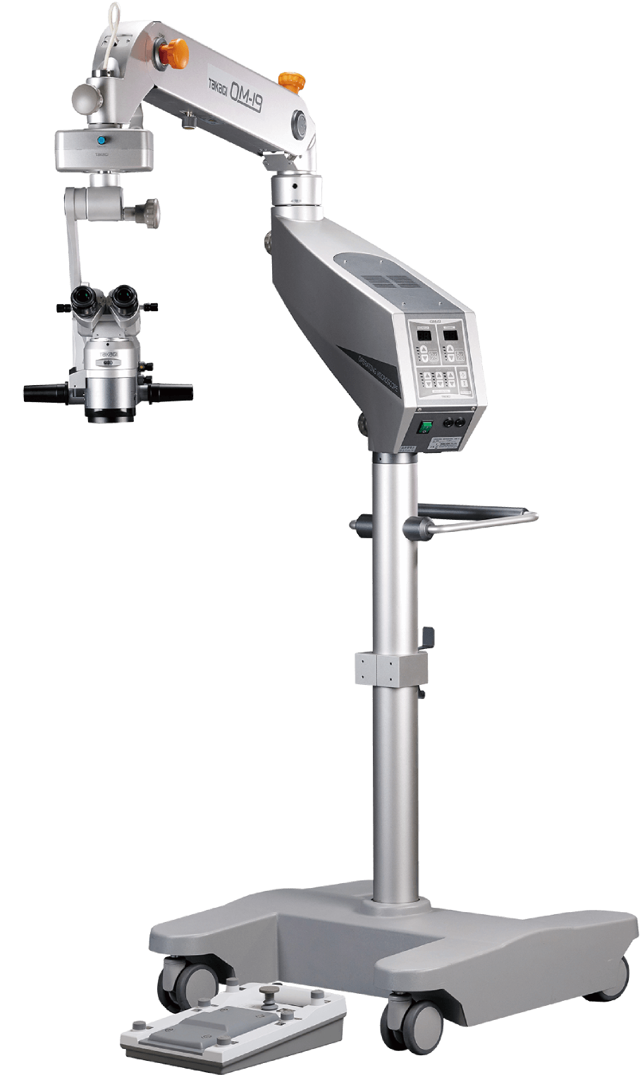
OM-19
Supports a wide range of surgeries, from cataract surgery to retinal vitrectomy.
Independent light sources provide a wide range of illumination.
Flexible customization with a rich variety of options.


All illuminations use high-luminance LED with excellent contrast color reproduction. A long service life of approximately 40,000 hours allows for maintenance-free illumination. Furthermore, the microscope is equipped with independent red reflex illumination LEDs for both the right eye and the left eye of the surgeon to provide both eyes with a stable red reflex image.
*LED operating life is defined as a state when light intensity drops to 70%. The operating life of the LED used in OM-19 is approximately 40,000 hours, although this is not guaranteed.
The focus, motorized zoom, X-Y speed adjustment, light intensity adjustment, and power switches are all integrated into the control panel to make operations more efficient. The light intensity can be adjusted using both the foot controller and the panel, and a staff member can also perform operations on the panel.
The individual light intensity values of the main illumination and red reflex illumination are displayed on the panel to enable smooth adjustments.
The main and red reflex illumination lights can be controlled independently, allowing flexible adjustment of visibility and contrast during surgery. By efficiently utilizing red reflex illumination reflected from the fundus, the system provides optimal brightness tailored to various surgical cases, supporting a comfortable and highly precise surgical environment.
The red reflex illumination is designed to efficiently capture light reflected from the fundus, enhancing visibility during surgery. It provides stable red reflex illumination even during procedures where red reflex is difficult to obtain—such as ultrasonic phacoemulsification and aspiration (PEA) or surgeries involving small pupils.
Using both the main and red reflex illumination together helps create a stereoscopic surgical view, while using red reflex illumination alone enhances the contrast of the lens. The system offers illumination optimized for visibility under a variety of surgical conditions.
The main illumination uses a high-luminance LED, supporting surgical observation with excellent contrast and color reproduction.
Additionally, the red reflex illumination mechanism can be retracted using the ON/OFF knob, allowing retinal vitreous surgeries to be performed using the system’s inherently bright optical system.
The main and red reflex illumination lights each use independent LED light sources, allowing individual brightness adjustments based on the condition of the surgical eye and the surgeon’s preference. Brightness levels for each illumination can be easily monitored and adjusted via numerical indicators on the control panel, enabling easy setting of intensity and balance.
This system allows the surgeon to enhance visibility in the surgical field and flexibly control contrast and reflections according to the situation. Its flexibility and operability support a more comfortable and efficient surgical environment suited to a variety of cases.
The combination of filters and high-performance optics supports procedures ranging from cataract to retinal vitreous surgeries. It delivers both high observation accuracy and gentleness to the eye, ensuring comfortable visibility during surgery.
The OM-19 is equipped with filters that take heat and wavelength characteristics into account. The heat-absorbing filter efficiently absorbs infrared light to reduce thermal load during surgery. The retina shield filter minimizes light exposure to the retina while maintaining clear visibility of the surgical field. Blue cut and blue collection filters properly control blue light to create an eye-friendly illumination environment.
These filters support both sustained observation accuracy and reduced strain on the eye during surgery.
The optical design supports use in retinal vitrectomy surgeries, and by attaching a wide-angle fundus observation device (sold separately), intraoperative observation is enabled. It is compatible with OCULUS’ BIOM, allowing selection according to the surgical procedure (*simultaneous use with the Coaxial Binocular Microscope O06-29SE is not possible when the BIOM is attached).
Additionally, the red reflex illumination mechanism can be retracted using the ON/OFF knob, allowing retinal vitreous surgeries to be performed using the system’s inherently bright optical system.
A large-diameter objective lens ensures a wide field of view and brightness, making the optical design suitable not only for cataract surgery but also for retinal vitreous surgery.
The optical system from the eyepiece to the objective lens is incorporates design features to suppress chromatic aberration, delivering a bright and sharp image with deep depth of focus.
The eyepieces feature high-eyepoint design lenses, supporting comfortable visibility during surgery.
The OM-19 is equipped with a multifunctional foot controller, an assistant microscope, and an adjustable tilt mechanism.
It provides the surgeon and assistant with optimal surgical field visibility and operability, supporting smooth and efficient surgery while reducing their workload and physical strain.
The foot controller features operation functions such as switching the mail and red reflex illumination on/off and illumination control, as well as controls that allow adjustment of focus, zoom, and X-Y directions. Additionally, the pedal layout can be customized to suit the surgeon’s operating style, enhancing ease of use.
The Coaxial Binocular Microscope (O06-29SE) provides a bright, wide field of view with deep depth of focus and a strong sense of stereopsis. It is also equipped with an independent focus adjustment function, offering optimal visibility for the assistant.
The Beam Splitter (O11-02, O11-03) and Camera Adapter (O08-11) enable video recording, supporting visualization and documentation of information.
The Monitor Arm (O06-29) integrates the LCD monitor with the surgical microscope, and the safety-designed Camera Control Rack (O06-30) supports a comfortable surgical environment.
A microscope head tilt mechanism suitable for glaucoma surgery is included, offering a wide range of motion of ±30°. The handle-based design allows quick and intuitive operation, enabling adjustment to the desired viewing angle.
This helps reduce the burden on the surgeon and contributes to a stable surgical environment.

Entry-level operating microscope suitable for anterior and periphery procedures. LED light source without changing a light bulb. Compact size microscope but high optical performance.

LED optimised system for outstanding clarity, can be configured in various specifications. Suitable for anterior and posterior surgical procedures.
 Home
Home Distributors Login