このホームページをご利用いただくための注意事項です。
このホームページは国内の医療従事者の方を対象に、タカギセイコーの眼科機器製品に関する情報を提供しています。
国外の医療関係者、一般の方への情報提供を目的としたものではありませんので、ご了承ください。
あなたは、日本国内の医療従事者ですか?
Operating Microscopes
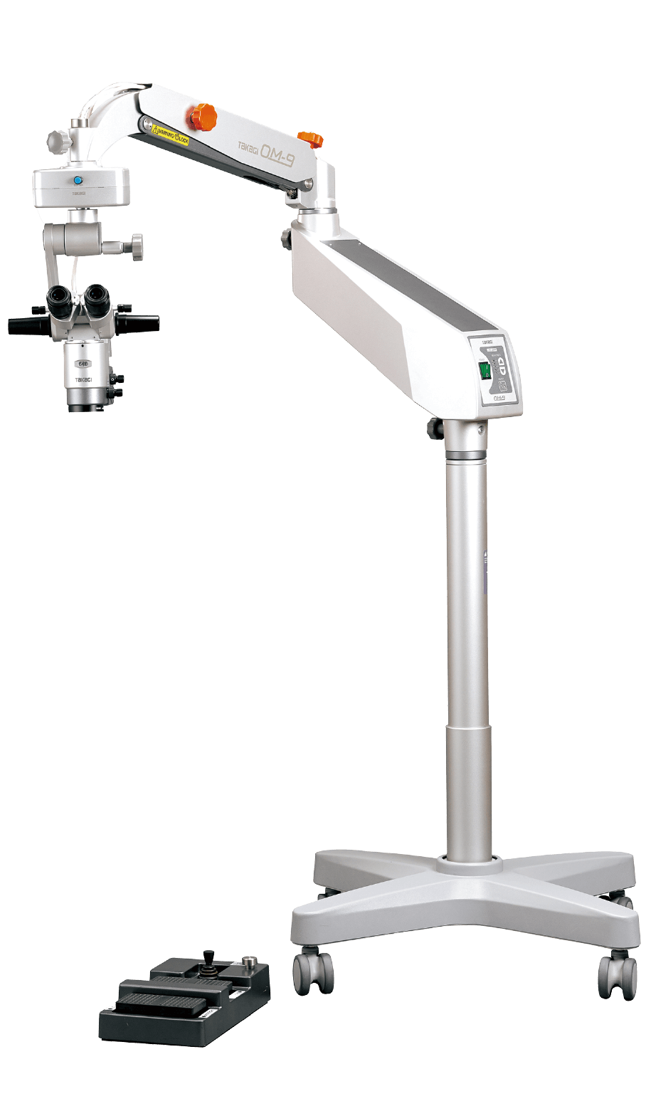
OM-9
Supports a wide range of operations, from cataract surgery and retinal vitrectomy to out-patient procedures.
Apochromatic lens and red reflex illumination deliver a clear field of view.
Flexible customization with a rich variety of options.


The optimized optical design and high-quality optical system maximize visibility during surgery and allow tiny tissues and structures to be identified clearly.
Combination of a red reflex illumination mechanism and high-luminance LED
The illumination system uses high-luminance LED light to focus on contrast and color reproduction. The red reflex illumination mechanism provides ample reflected light from the fundus, resulting in high-quality red reflex and 3D observation images, which are essential for cataract surgery.
Three configuration options are available according to need
There are three types of specification configurations for the OM-9 depending on the combination of coupling, microscope and binocular tubes, making it possible to configure the optimal device for your specific needs.
*Configurations other than the three basic types listed above may be possible. For further details, please contact our sales department.
The high-intensity LED, red reflex illumination mechanism, and deep depth-of-focus lens enhance visibility during surgery and color reproducibility, contributing to enhanced surgical safety and accuracy.
The system is equipped with a red reflex illumination mechanism using a high-intensity LED so that when red reflex illumination is turned ON, it switches to a structure where light is emitted from two mirrors. This makes it easier to obtain reflected light from the fundus, enabling red reflex observation required for cataract surgery and similar procedures.
This design ensures a naturally bright and three-dimensional surgical field, making it easier to grasp the condition of the affected area.
LEDs feature high brightness and long lifespan for enabling stable illumination over a long period of time. The OM-9 incorporates a dedicated filter designed to ensure color reproduction and visibility during observation while taking advantage of LED characteristics. Special attention has been paid to the blue wavelength to enhance color contrast and improve visibility of the target tissues.
The F = 200 mm objective lens provides deep depth of focus, making it easy to keep the entire surgical field in focus during surgeries. This allows continued observation with minimal focus deviation, even when the target area moves slightly.
Also, the OM-9 uses an apochromatic lens for correcting chromatic aberration across red, blue, and green wavelengths, reducing color fringing and supporting observation with more natural coloration.
With a variety of combinations including binocular tubes and magnification adjustment, the system provides comfortable operation and an efficient observation environment tailored to the surgeon’s style.
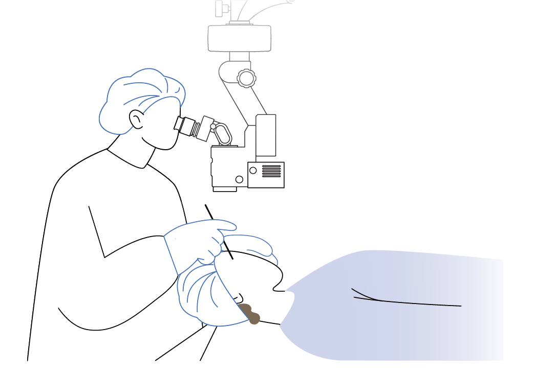
The XZ Configuration (motorized zoom microscope specification) features a tiltable binocular tube adjustable by more than 90° for enabling observation in a comfortable posture. Fine adjustment of X-Y movement is also possible, supporting surgical field alignment. Magnification can be continuously varied using the motorized zoom and is operable using the foot controller.
This enables smooth switching of the surgical field and adjustment of the magnification during surgery without interrupting the workflow, allowing full concentration. This configuration provides a balance of comfort and operability.
The XE Configuration (motorized 5-step magnification microscope specification) features a fixed binocular tube angled at 45° and allows fine adjustment of X-Y movement. The motorized 5-step magnification function enables magnification changes using the foot controller.
This allows the surgeon to smoothly change magnification, supporting a comfortable observation environment. This configuration provides a balance between cost and usability.
The SM Configuration (manual 5-step magnification microscope specification) provides the same optical performance as the XZ and XE Configurations and is suitable for use in outpatient procedures. Focusing on essential functions, magnification is adjusted manually. This configuration’s simple operability allows for proper setting of the required observation magnification, supporting ease of use in everyday procedural environments.
Foot controllers, assistant microscopes, and a microscope head tilt mechanism enable customization of operation and layout based on configuration, providing a comfortable surgical view and operating environment.
In all microscope configurations, the foot controller can be used for ON/OFF control of illumination and focus adjustment. In the XZ and XE Configurations, the foot controller also supports magnification switching and X-Y movement. Furthermore, the pedal layout for focus and magnification adjustment can be customized to match the surgeon’s preference for enhanced operability.
It also features waterproof and dustproof performance (IPX6), ensuring safe use even in environments where liquids may splash.
*Image with LCD monitor and monitor arm O06-22 attached.
Assistant Microscopes (O06-19SE, O06-20SE) share the same field of view with the surgeon, facilitating smooth surgical collaboration.
Beam Splitters (O11-03) and Camera Adapters (O08-11, O08-22) support visualization and recording of information.
The Monitor Arm (O06-22) allows integration of the LCD monitor with the surgical microscope, and the safety-designed Camera Control Rack (O06-23) supports a comfortable surgical environment.
A microscope head tilt mechanism suitable for glaucoma surgery is included, offering a wide range of motion of ±30°. The handle-based design allows quick and intuitive operation, enabling adjustment to the desired viewing angle.
This helps reduce the burden on the surgeon and contributes to a stable surgical environment.

Entry-level operating microscope suitable for anterior and periphery procedures. LED light source without changing a light bulb. Compact size microscope but high optical performance.

The world's first operating microscope with independent light intensity adjustment for coaxial and red-reflex illumination. With X-Y coupling, zoom magnification, tiltable eyepieces and rotating coaxial stereoscopic assistant microscope. Suitable for anterior and posterior surgical procedures.
 Home
Home Distributors Login