Serving Your Vision
このホームページをご利用いただくための注意事項です。
このホームページは国内の医療従事者の方を対象に、タカギセイコーの眼科機器製品に関する情報を提供しています。
国外の医療関係者、一般の方への情報提供を目的としたものではありませんので、ご了承ください。
あなたは、日本国内の医療従事者ですか?
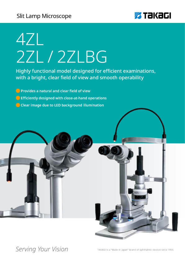
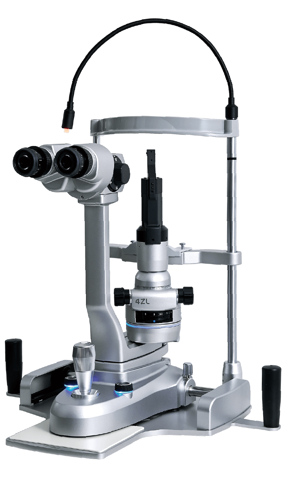
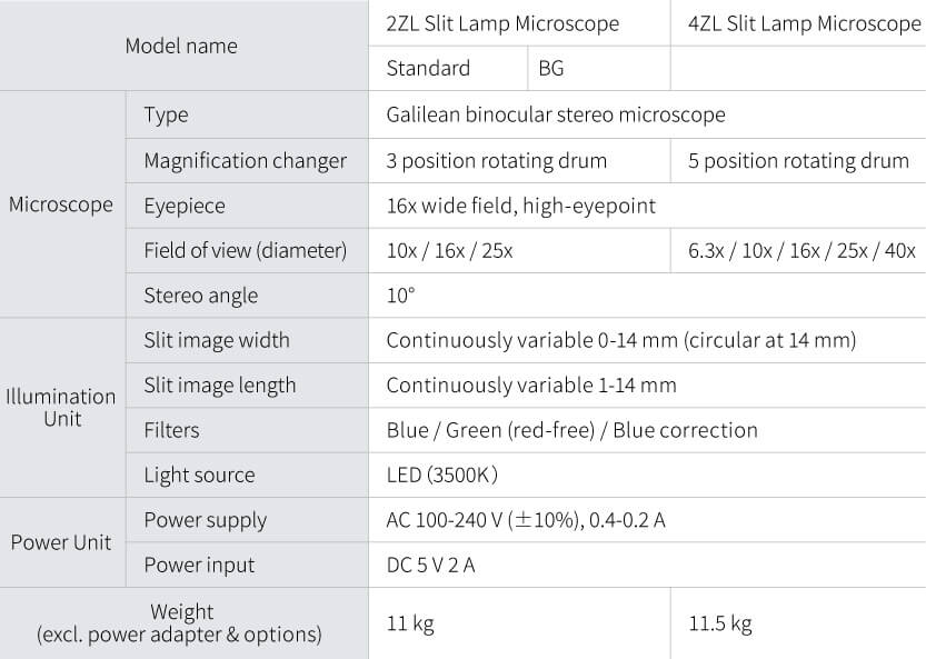
Slit Lamp Microscopes
4ZL
●Provides a natural and clear field of view
●Efficiently designed with close-at-hand operations
●Clear image due to LED background illumination
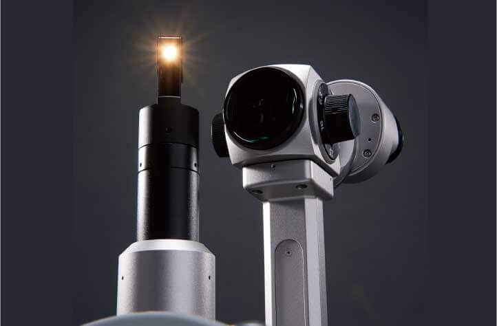
A high-luminance LED light source is used to enable affected areas to be observed clearly and in detail for more accurate diagnosis. Color and light unevenness are reduced to provide a natural and clear field of view.
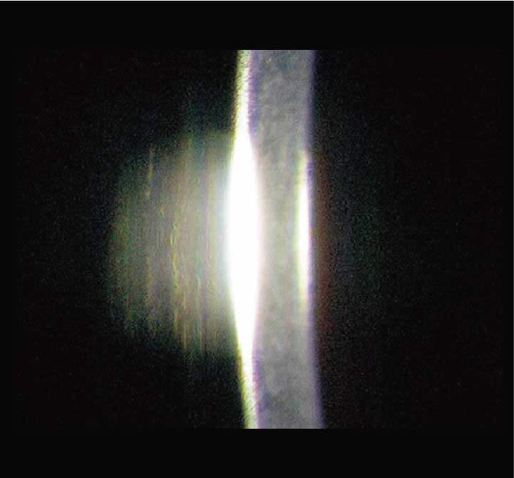
The inter-optical path distance of 22 mm and the sharp slit illumination that uses a high-luminance LED with a 3500 K color temperature
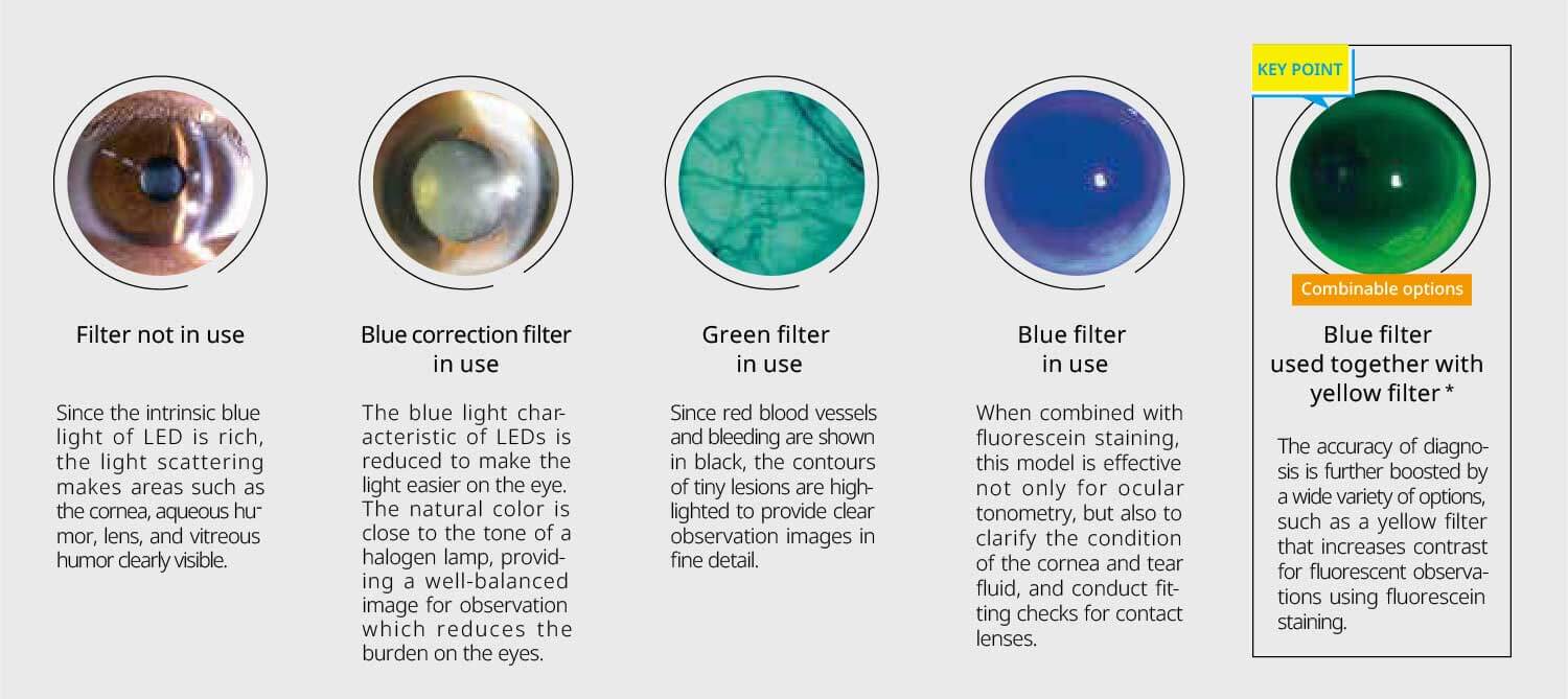
*Photo provided by: Dr. Toru Noda, Department of Ophthalmology, NHO Tokyo Medical Center
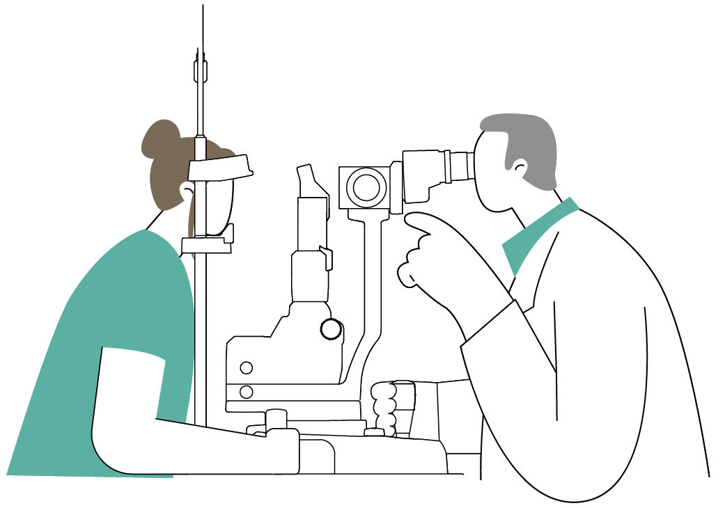
To reduce the burden on doctors during long examinations and minimize fatigue, the design focuses on ease of use, from the size and positioning of the operating parts to their operational feel.
This model uses an energy-saving LED light source which provides stable brightness over a long service life.
This allows a design with built-in cables and reduces heat generation, contributing to enhanced patient safety.
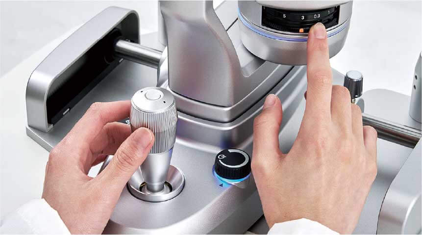
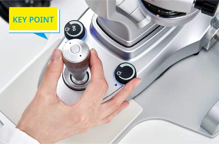
The design consists of parts such as a large-diameter dimmer knob arranged functionally around the joystick for easy operation with one hand.
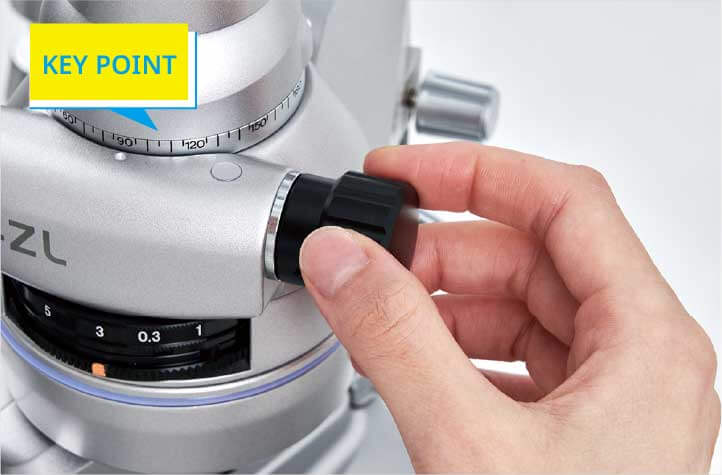
The size and feel of the large-diameter control knob are optimally designed for comfortable operation.
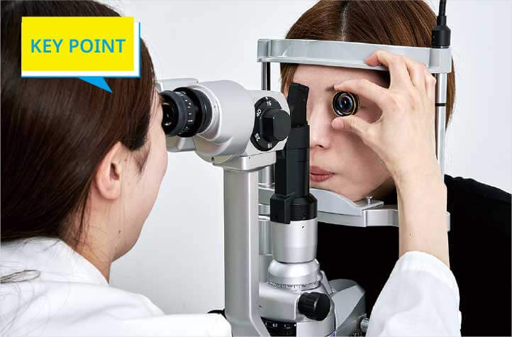
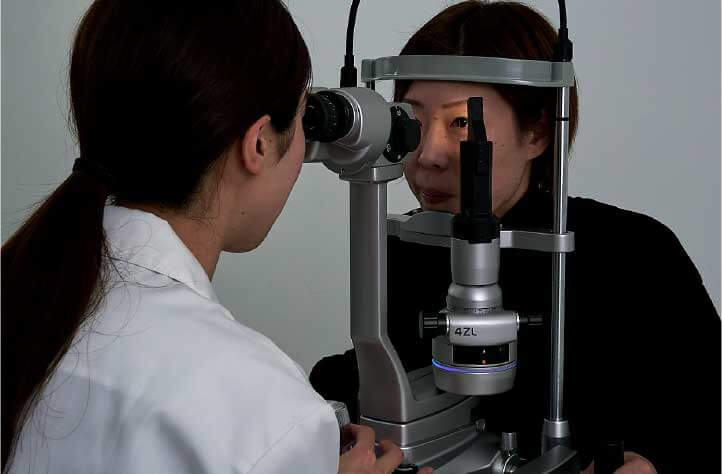
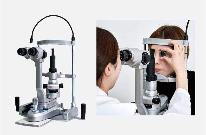
The front lens can be held in a stable position using the finger rest function of the forehead rest.
This makes observations easier and reduces the burden on the doctor’s arms, while also minimizing contact with patients.
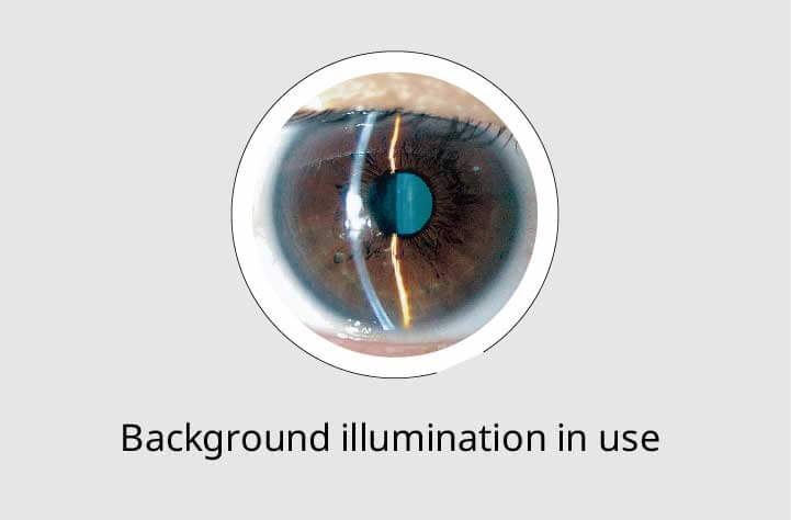
The background illumination uses an LED with the same color temperature as the slit illumination to obtain a clear image without impairing the color balance.
This makes the entire eye easy to view, allowing the position and condition of affected areas to be communicated intuitively to patients.
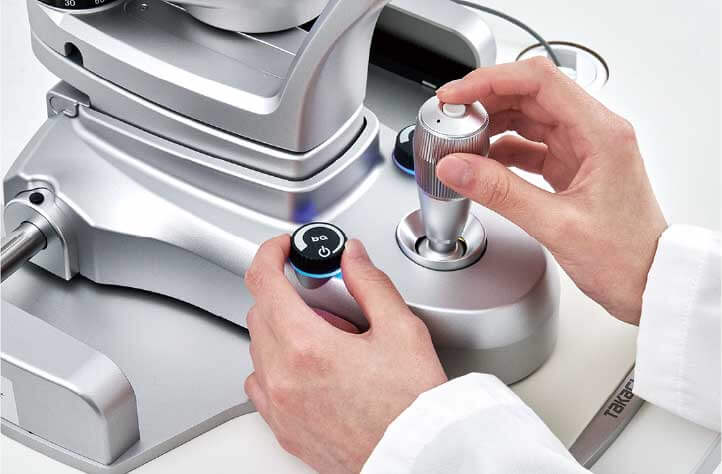
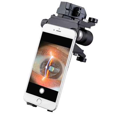
Press a single button to capture images while operating the joystick.
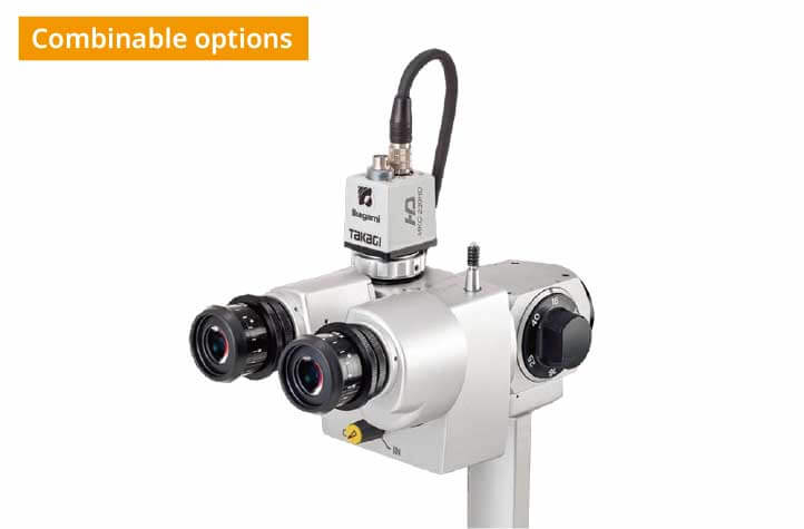
A camera for capturing images can be connected by installing a combination adapter or camera adapter made by TAKAGI.
The high-luminance LED delivers natural color and clear imaging, contributing to comfortable examinations and enhanced diagnostic accuracy.
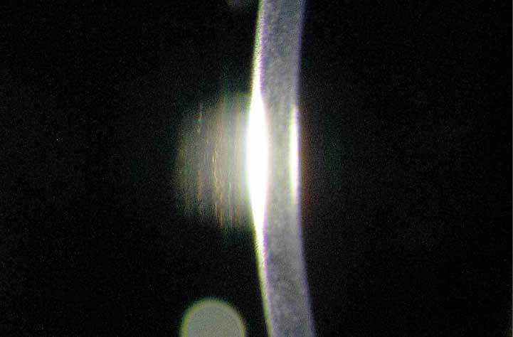
A single-element, high-luminance LED provides natural color and a uniform slit beam. Rich light scatter enhances tissue contrast, enabling clear observation of the cornea, aqueous humor, lens, and vitreous humor.
The microscope features converging binocular tubes with a 6° convergence angle, making fusion easier and providing a natural sense of depth.
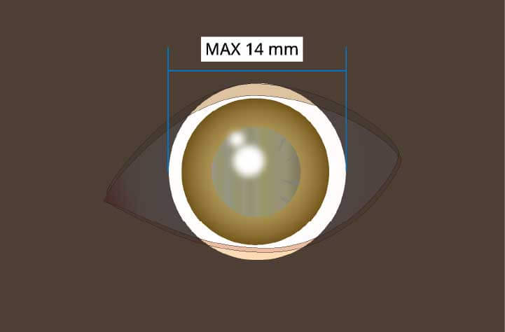
A wide 14 mm illumination field diameter is used to provide sufficient coverage for the cornea, which is approximately 11 mm in diameter.
Even if the eye moves slightly, the entire cornea remains within the illumination field, allowing stable observation.
By enhancing patient safety and reducing the burden on physicians, this system supports comfortable, smooth, and efficient examinations.
The power and connection cables are housed internally within the main unit, minimizing their external exposure. This reduces the risk of contact with the patient, enhances cleanliness, and ensures smooth handling and ease of operation.
The finger rest on the forehead support improves stability when using a front lens, reducing strain on the physician’s arm while also minimizing contact with the patient.
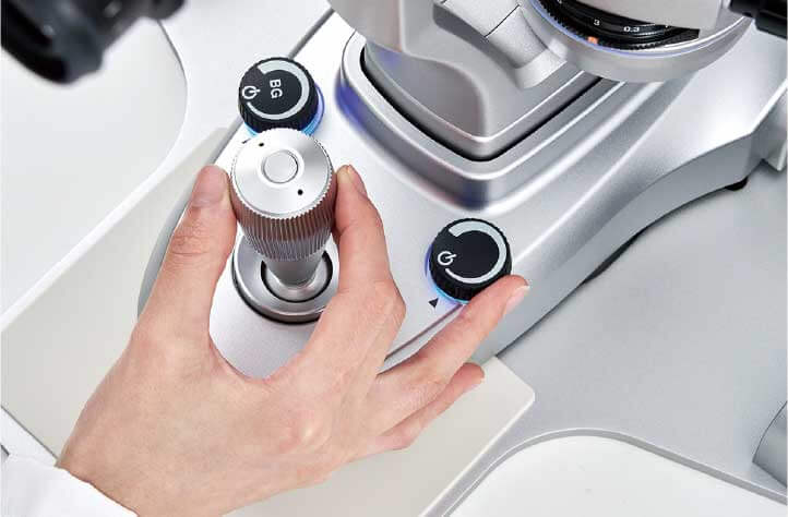
The slit illumination dimming knob and background illumination dimming knob, positioned around the joystick, are thoughtfully designed for comfort in size and tactile feel. Their intuitive one-handed operation allows adjustments while looking through the microscope. Additionally, the trigger button on top of the joystick enables image capture without releasing the joystick.
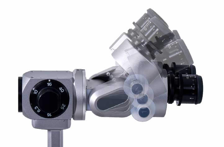
*Image with Tiltable Binocular Tube O12-20 attached to 700GL.
Using optional components such as the Tiltable Binocular Tube O12-20 or the 13-degree Tilt Adapter S06-59 helps reduce tension in the neck and shoulders, supporting a better work environment with more comfortable posture during examinations.
The use of high-eyepoint eyepieces makes observation easy even while wearing glasses.
There is no need to remove glasses during observation, enhancing convenience in the examination environment.
The built-in background illumination optimize the imaging environment. Support for a wide range of camera connections enables high-precision recording of examination findings.
The background illumination is built into the main unit. Because it is not an external lighting unit, there is no concern about the physician’s hand interfering with protruding parts of the illumination.
The background LED uses the same specifications as the main illumination. Although it shares the same color temperature, it operates independently from the main light, allowing both light sources to be adjusted freely. This enables well-balanced image capture with consistent brightness and color tone.
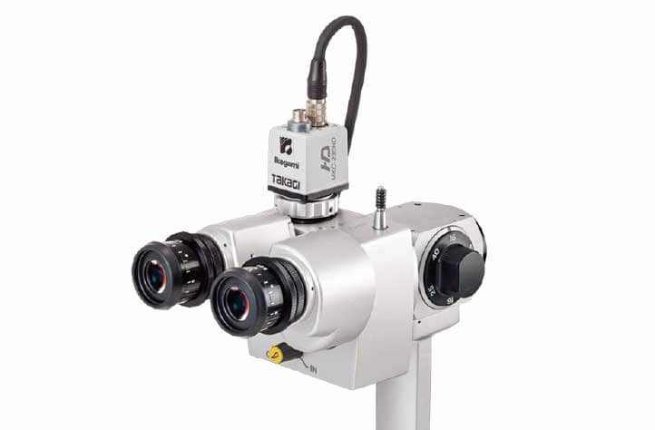
*Image with Digital Camera Adapter TD-2 attached to 700GL.
In addition to shooting with a mounted TAKAGI digital camera, a single-lens reflex camera can also be mounted using a camera adapter.
Image capture can be easily performed using the trigger button located on top of the joystick.
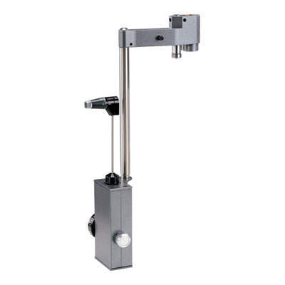
R-Type Compact Applanation Tonometer with option to be permanently fixed to slit lamp with observation from the left eye
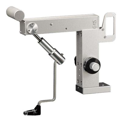
Z-type applanation tonometer exclusively for use with 2ZL/4ZL
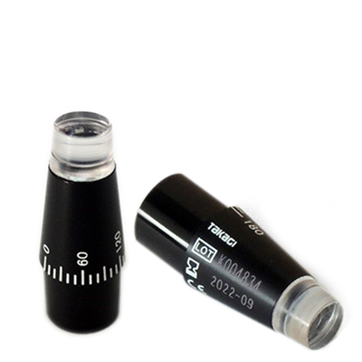
Reusable Measuring Prism for AT-1 Applanation tonometer
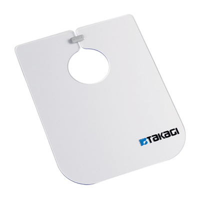
Infection control item
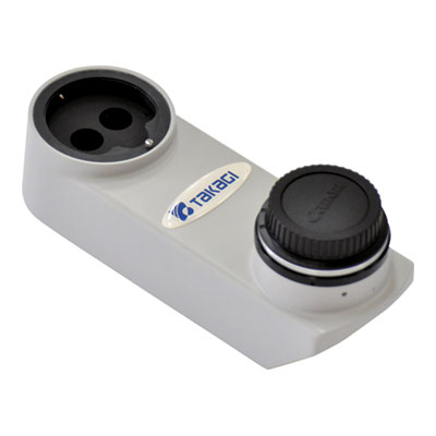
Beam Splitter with integrated yellow filter, attachable to Canon EOS digital camera
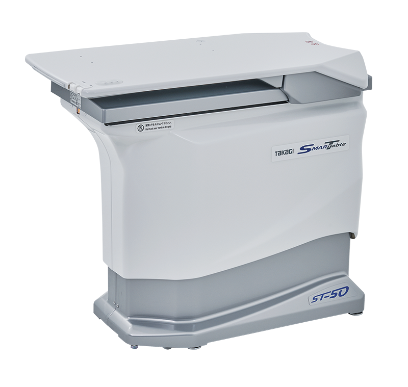
Wheelchair compatible motorised table with fingertip swipe movement function , LED back-lit switches and manual slide mechanism
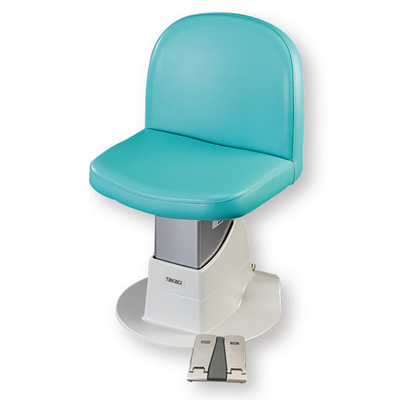
Motorised Patient Chair
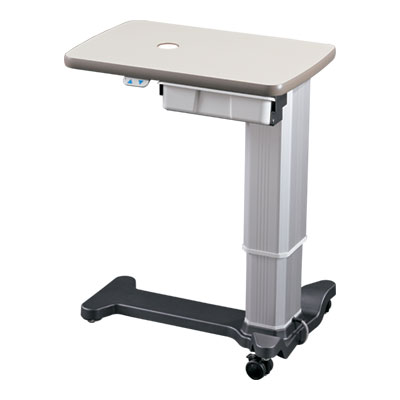
Wheelchair friendly motorised table
Equipped with a 12.4-megapixel high-resolution image sensor and an Auto Exposure function that enabling you to capture images optimised to lighting conditions
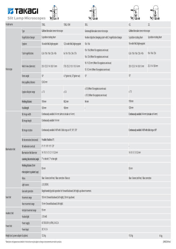
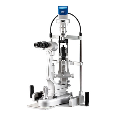
5x step Galilean type, converging optics, upper LED illumination, and built-in background illumination
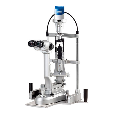
5x step Galilean type, converging optics, upper LED illumination, built in background illumination and wide angle fundus observation
2x step Greenough type with converging optics and upper LED illumination
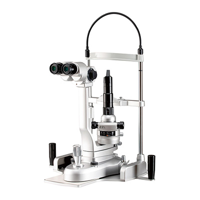
3x step Galilean type with converging optics and lower LED illumination
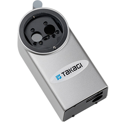
Portable, LED lighting, anterior segment observation, excellent operability
R-Type Compact Applanation Tonometer with option to be permanently fixed to slit lamp with observation from the left eye
Z-type applanation tonometer exclusively for use with 2ZL/4ZL
Reusable Measuring Prism for AT-1 Applanation tonometer
Infection control item
 Home
Home Distributors Login