Serving Your Vision
このホームページをご利用いただくための注意事項です。
このホームページは国内の医療従事者の方を対象に、タカギセイコーの眼科機器製品に関する情報を提供しています。
国外の医療関係者、一般の方への情報提供を目的としたものではありませんので、ご了承ください。
あなたは、日本国内の医療従事者ですか?
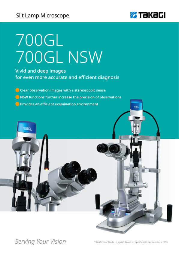
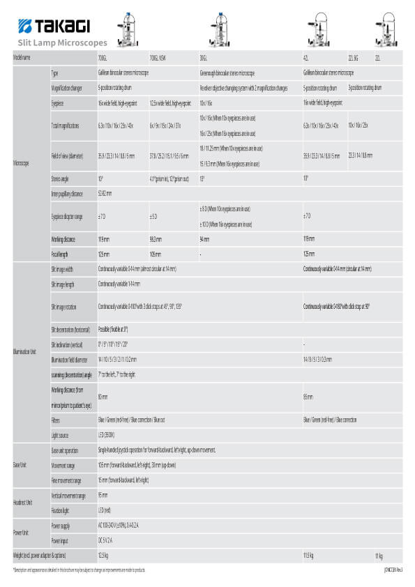
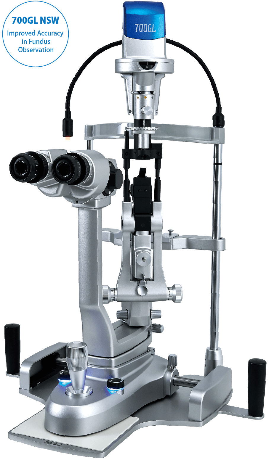
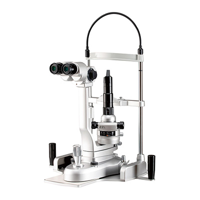
Slit Lamp Microscopes
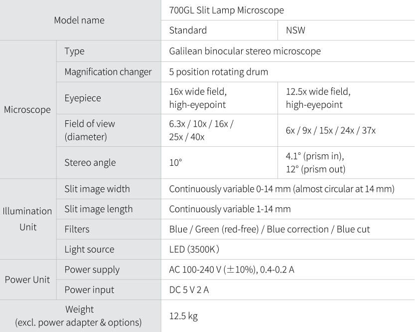
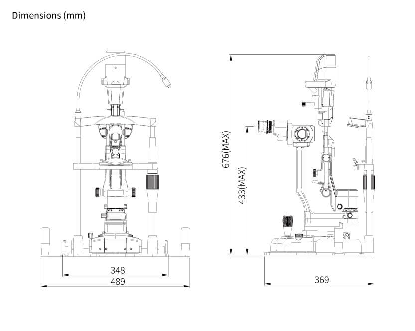
700GL NSW
●Clear observation images with a stereoscopic sense
●NSW functions further increase the precision of observations
●Provides an efficient examination environment
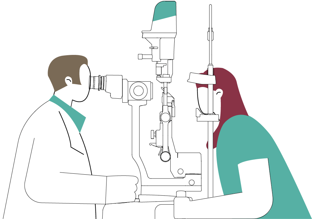
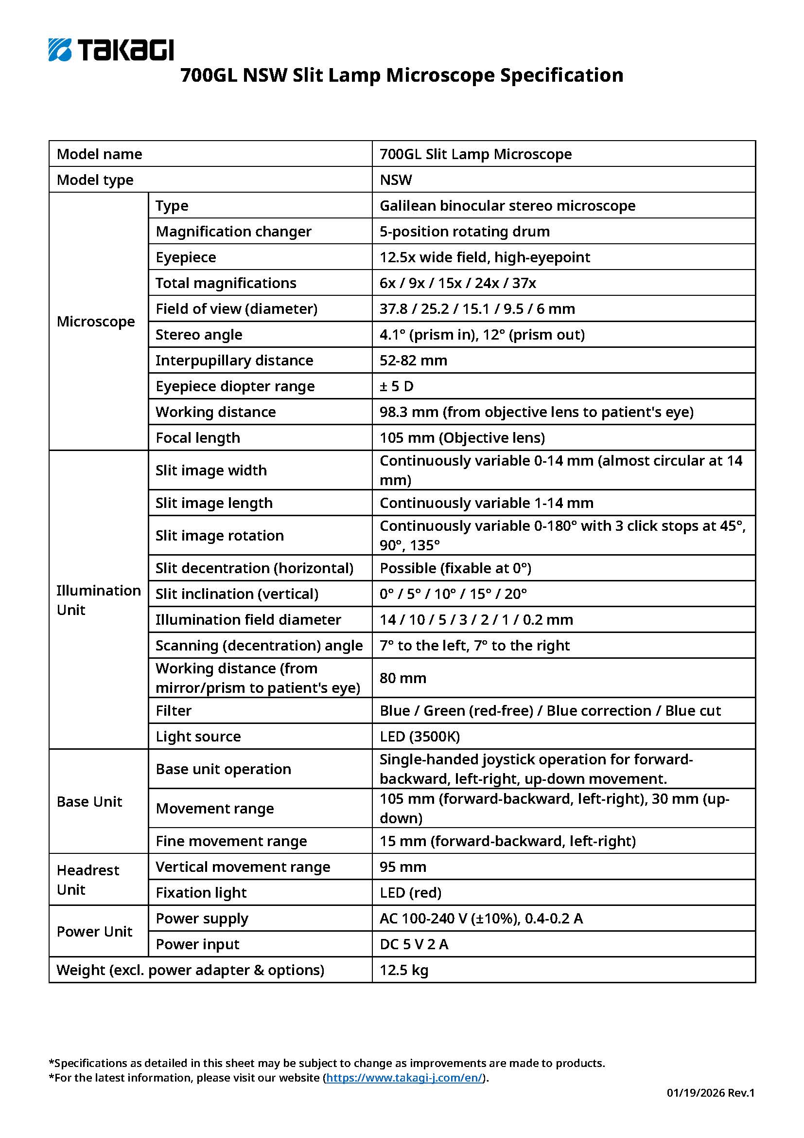
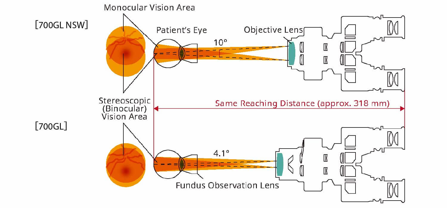
In contrast to the 10° stereo angle of the 700GL, an angle of 4.1° is adopted for the NSW, ensuring a wider stereoscopic vision area. This makes it easier to observe the fundus in detail for highly accurate diagnosis.
*The image is for illustrative purposes only.
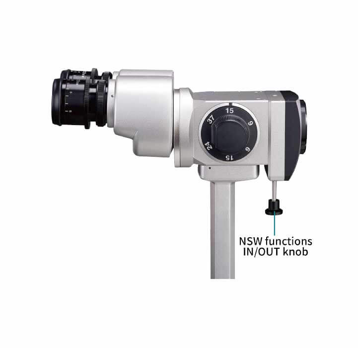
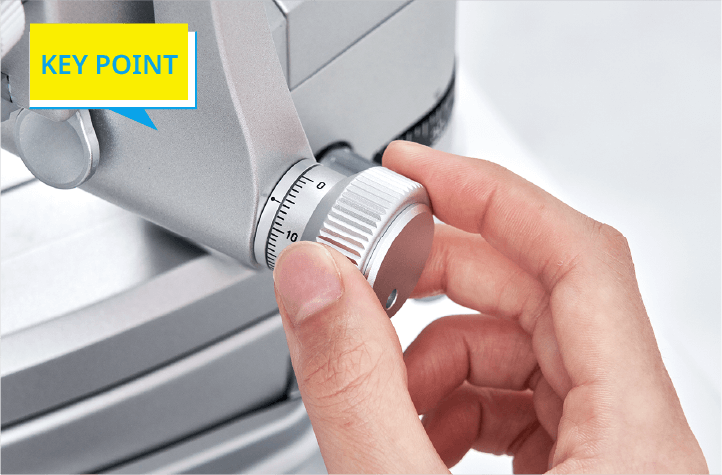
Switching to the NSW functions is easy. Simply press the knob at the bottom of the objective lens.
The inter-optical path distance of 22 mm and the sharp slit illumination that uses a high-luminance LED with a 3500 K color temperature produce a clear observation image with a stereoscopic sense from the anterior eye parts to the fundus. Lesions and foreign matter can be identified clearly and in detail, supporting more accurate diagnosis.
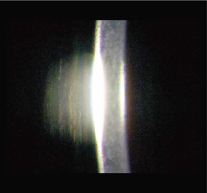
The inter-optical path distance of 22 mm and the sharp slit illumination that uses a high-luminance LED with a 3500 K color temperature
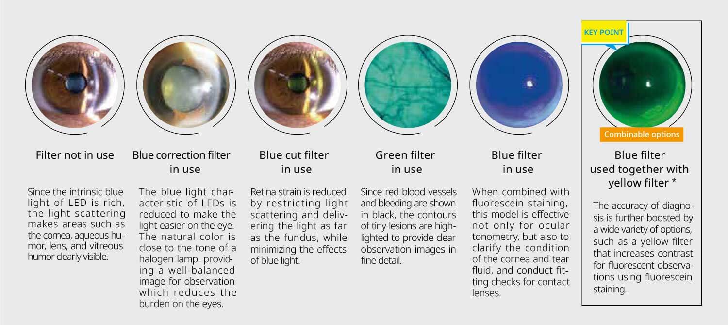
*Photo provided by: Dr. Toru Noda, Department of Ophthalmology, NHO Tokyo Medical Center
To reduce the burden on doctors during long examinations and minimize fatigue, the design focuses on ease of use, from the size and positioning of the operating parts to their operational feel. This model uses an energy-saving LED light source which provides stable brightness over a long service life. This allows a design with built-in cables and reduces heat generation, contributing to enhanced patient safety.
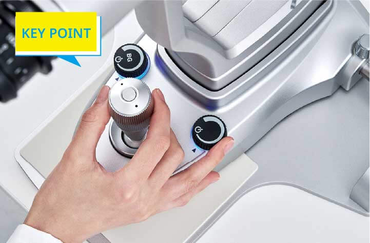
The design consists of parts such as a large-diameter dimmer knob arranged functionally around the joystick for easy operation with one hand.
The size and feel of the large-diameter control knob are optimally designed for comfortable operation.
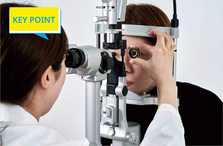
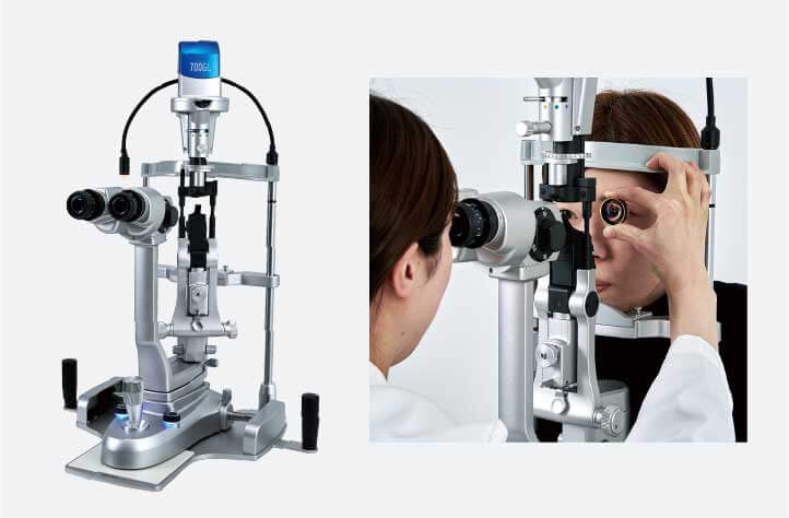
The front lens can be held in a stable position using the finger rest function of the forehead rest. This makes observations easier and reduces the burden on the doctor’s arms, while also minimizing contact with patients.
Designed with prisms that expand the binocular viewing range, it provides clear stereoscopic images. This enables high-precision binocular observation, enhancing the quality of fundus examinations.
Prisms that adjust the stereo angle expand the binocular viewing range, increasing the stereoscopic viewing area to approximately 133% compared to the 700GL model. This allows clear and detailed stereoscopic visualization of fine fundus structures. Additionally, the smaller stereo angle is advantageous for stereoscopic observation in patients with small pupils. By adopting a design that minimizes the depth of the device, it maintains a reaching distance equivalent to the 700GL, ensuring that the distance to the patient remains short and the operator can easily reach the patient-supporting a comfortable examination environment.
By enhancing patient safety and reducing the burden on physicians, this system supports comfortable, smooth, and efficient examinations.
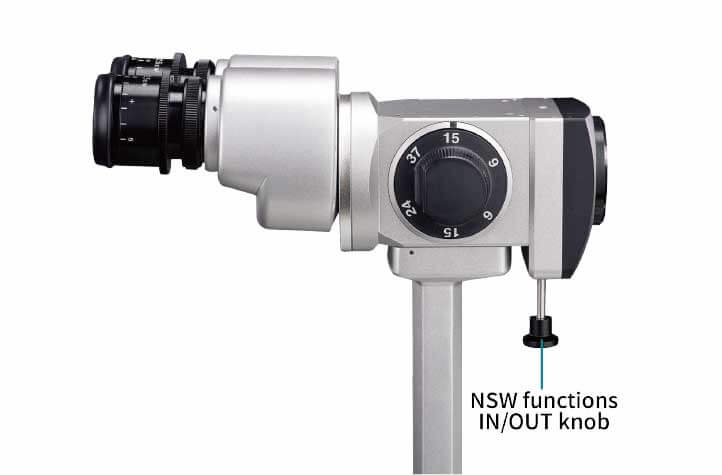
During fundus observation, pressing the knob upward activates the prism. The distance between the physician and the patient remains unchanged from normal observation, maintaining a smooth examination flow and a design that considers operational efficiency.
The power and connection cables are housed internally within the main unit, minimizing their external exposure. This reduces the risk of contact with the patient, enhances cleanliness, and ensures smooth handling and ease of operation.
The finger rest on the forehead support improves stability when using a front lens, reducing strain on the physician’s arm while also minimizing contact with the patient.
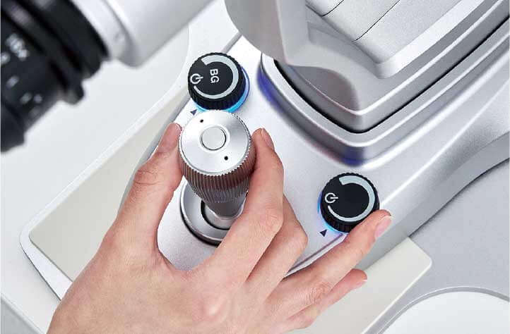
The slit illumination control knob and background illumination control knob, positioned around the joystick, are thoughtfully designed for comfort in size and tactile feel. Their intuitive one-handed operation allows adjustments while looking through the microscope. Additionally, the trigger button on top of the joystick enables image capture without releasing the joystick.
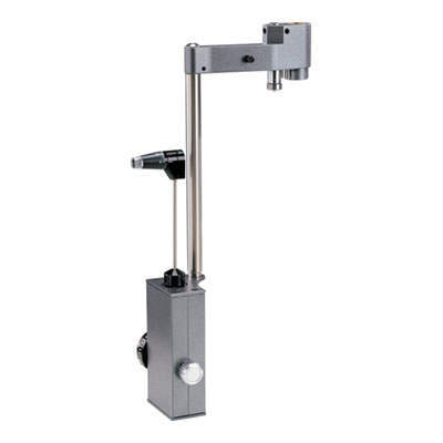
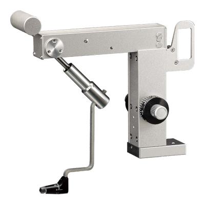
R-Type Compact Applanation Tonometer with option to be permanently fixed to slit lamp with observation from the left eye
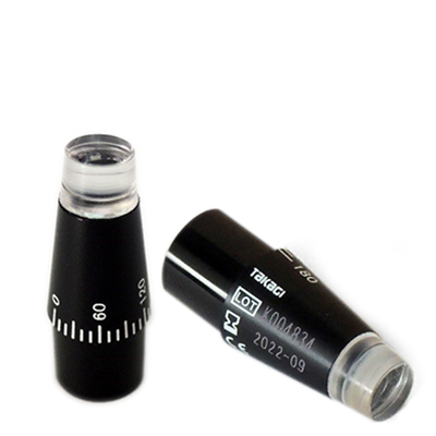
Reusable Measuring Prism for AT-1 Applanation tonometer
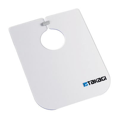
Infection control item
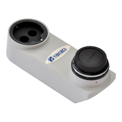
Beam Splitter with integrated yellow filter, attachable to Canon EOS digital camera
Wheelchair compatible motorised table with fingertip swipe movement function , LED back-lit switches and manual slide mechanism
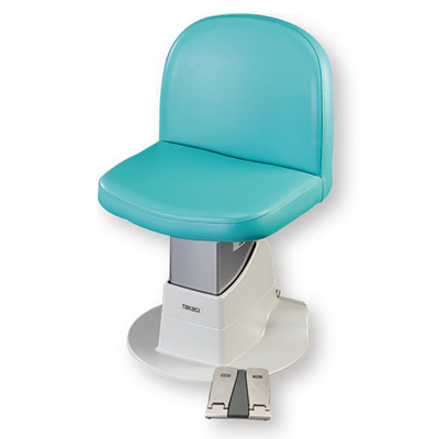
Motorised Patient Chair
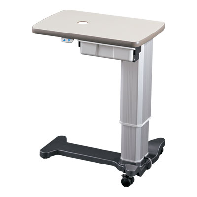
Wheelchair friendly motorised table
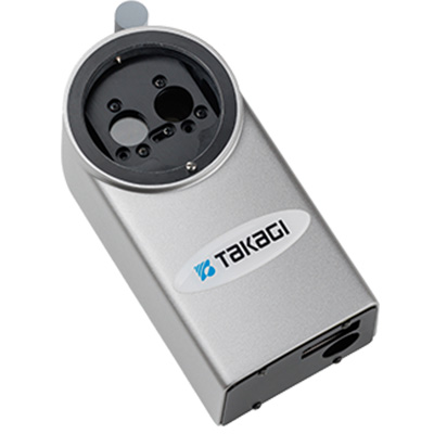
Equipped with a 12.4-megapixel high-resolution image sensor and an Auto Exposure function that enabling you to capture images optimised to lighting conditions
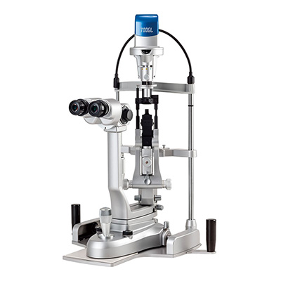
5x step Galilean type, converging optics, upper LED illumination, and built-in background illumination
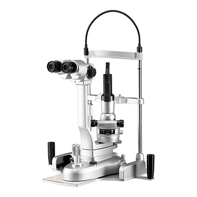
2x step Greenough type with converging optics and upper LED illumination
5x step Galilean type, converging optics, lower LED illumination, and built in background illumination
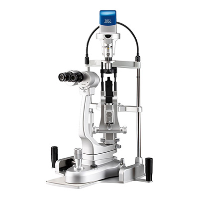
3x step Galilean type with converging optics and lower LED illumination
3x step Galilean type with converging optics and lower LED illumination
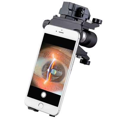
Portable, LED lighting, anterior segment observation, excellent operability
R-Type Compact Applanation Tonometer with option to be permanently fixed to slit lamp with observation from the left eye
Z-type applanation tonometer exclusively for use with 2ZL/4ZL
Reusable Measuring Prism for AT-1 Applanation tonometer
Infection control item
 Home
Home Distributors Login